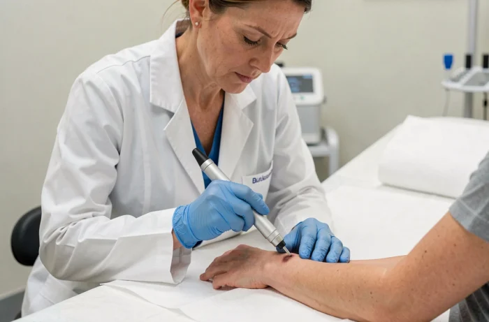
Skin cancer encompasses several distinct pathologies, with visual forms and prognosis varying considerably. Carcinomas (basal cell and squamous cell) account for the vast majority of cases, while melanoma, which is rarer, accounts for most of the mortality. Identifying a suspicious lesion relies on specific criteria, but also on regular monitoring that the population still applies too little.
Increasing UV Dose in Europe: A Worsening Risk Factor
Analyses covering the period from 2013 to 2022 show a significant increase in the annual UV dose received at many European stations, with a particularly clear rise in Central and Western Europe. This environmental observation directly alters the assessment of population risk.
The majority of skin cancers are attributable to ultraviolet radiation, whether natural or artificial. With the measured increase in exposure, dermatologists expect a rise in the number of diagnoses in the coming years, including among populations that consider themselves to be minimally exposed.
Preventive behaviors remain insufficient. A survey conducted in 2025 in the UK reveals that nearly half of adults reported having suffered a sunburn in the previous year, and a notable proportion takes no regular protective measures. These data, although from a country not known for its sunshine, illustrate a persistent gap between awareness of risk and actual practices.
To familiarize oneself with the visual aspect of suspicious skin lesions, consulting photos of skin cancer helps better understand what to look for during a self-examination.

Basal Cell and Squamous Cell Carcinoma: What Appearance Doesn’t Always Reveal
The basal cell carcinoma is the most common form. It often presents as a small, pearly, pinkish, sometimes translucent bump located on sun-exposed areas: face, ears, scalp, neck. The lesion may ulcerate, bleed intermittently, and then partially heal before reappearing.
Squamous cell carcinoma takes on different appearances. It can resemble a rough patch, a persistent crust, or a firm nodule that grows. It also appears on photo-exposed areas but can develop on old scars or areas of chronic inflammation.
What complicates early detection is that these two types of carcinomas sometimes mimic benign lesions. A small wound that does not heal within a few weeks, a recurring crust in the same spot, or a seemingly innocuous bump deserves a dermatological opinion. Any lesion that does not heal within three to four weeks should be examined.
Actinic Keratosis: The Precancerous Lesion to Monitor
Actinic keratosis appears as a scaly, dry, rough-to-the-touch spot, ranging in color from pink to brownish. It is not cancer in itself, but a proportion of these lesions can progress to squamous cell carcinoma if left untreated. Their presence indicates a cumulative sun exposure and warrants close monitoring.
Cutaneous Melanoma: The ABCDE Rule and Its Limitations
Melanoma develops from melanocytes, the pigment-producing cells. It can appear on an existing mole or on healthy skin, including areas rarely exposed to the sun (soles of the feet, nails, mucous membranes).
The ABCDE rule remains the reference tool for self-monitoring:
- Asymmetry: the lesion does not have a regular shape, one half does not resemble the other
- Irregular borders: the edges are scalloped, poorly defined, or blurry
- Heterogeneous color: presence of multiple shades (brown, black, red, white, blue) within the same lesion
- Diameter larger than that of a pencil eraser, although smaller melanomas exist
- Evolution: any change in size, shape, color, or thickness over time
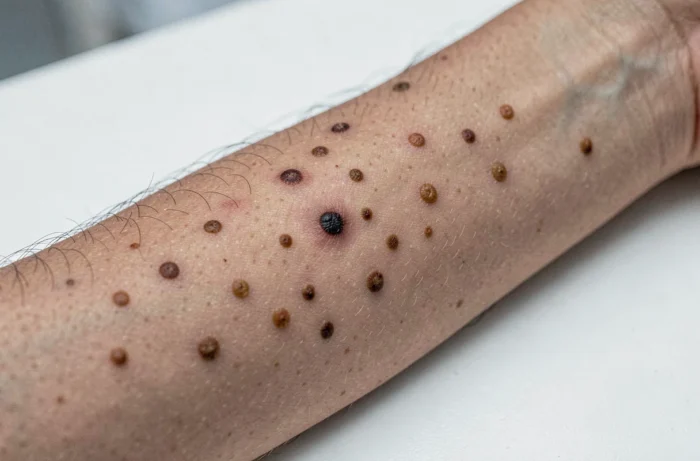
However, this grid has known limitations. Nodular melanomas, for example, do not always meet the classic criteria: they sometimes present as a symmetrical nodule, of uniform color (pink, red, or flesh-colored), that grows rapidly. Available data do not allow for concluding that a self-examination based solely on the ABCDE rule is sufficient to detect all melanoma subtypes.
Merkel Cell Carcinoma: Rare but Aggressive
Merkel cell carcinoma is a rare skin cancer, most often presenting as a firm, painless nodule, red or purplish in color, growing rapidly. It primarily affects older individuals and immunocompromised subjects. Its diagnosis is often delayed, as the lesion is easily confused with a benign cyst.
Skin Cancer Prevention: What Has Changed in Sun Regulation
Sun protection remains the primary lever for preventing skin cancers. However, the regulatory framework for sunscreen products is evolving. The European Commission has removed the substance 4-MBC from the list of authorized UV filters via regulation (EU) 2024/996, with a phased implementation starting in 2025-2026. This withdrawal directly affects certain formulations of sunscreens available in Europe.
At the same time, new in vitro and hybrid methods for measuring SPF were introduced in 2024, aimed at reducing reliance on human skin testing. This technical evolution could ultimately change the way protection indices are established and compared between products.
Beyond the choice of sunscreen, prevention relies on complementary actions:
- Avoid direct exposure between 12 PM and 4 PM, when UV rays are most intense
- Wear covering clothing, a wide-brimmed hat, and filtering sunglasses
- Reapply sunscreen every two hours and after each swim
- Never use a tanning bed, classified as a definite carcinogen
An annual dermatological examination is still recommended, especially for fair-skinned individuals, those with a family history of melanoma, or a high number of moles. Detecting a suspicious lesion at an early stage significantly improves the prognosis, regardless of the type of skin cancer involved.